


pyHREM-DIMA
(High Resolution Electron Microscopy Digital Image Matching Analysis)
last update: 31.08.2023
pyHREM-DIMA-setup.exe – 
_
pyHREM-DIMA Demo (Aug 31, 2023)
_
- A significant effort has been invested during the last few decades into developing new methods to allow quantitative analysis of HR-S/TEM images with the goal of determination of the exact positions of atoms. One approach (described in Figure 1), which has been extensively used by Mobüs et al. [1] for the study of interfaces [2], is based on iterative digital image matching (IDIM) of various model structures by comparison of the experimental image contrast to the one obtained from multi-slice image calculations [3].
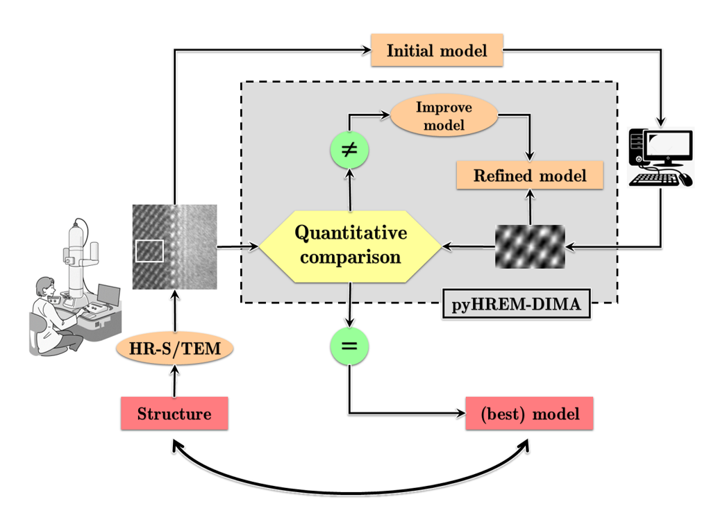
Figure 1 : A schematic representation of the concept of iterative digital image matching (IDIM).
- The software program, named High Resolution Electron Microscopy Digital Image Matching Analysis (pyHREM-DIMA), is basically based on the same concept as IDIM. So far, most programs which use the IDIM approach and implement the image matching in a fully automated process frequently fail and may produce inaccurate results. The main reason for the failure of these algorithms is that the fully automated iterative matching process frequently gets trapped in a local optimum instead of locating the true global optimum.
- The new approach which was implemented in pyHREM-DIMA combines an interactive matching process done by the user controlling the very intuitive graphical user interface (GUI), and a fully quantitative process of comparison between the experimental and simulated micrographs. The fully quantitative results which can be obtained by this approach allow determining very precisely the exact conditions at which the experimental data was acquired and ultimately the positions of the projected atomic columns in the specimen.
- This open-source software program was developed using QT and Python (see Figure 2 for the interactive GUI) and is available as a standalone application for Windows (tested under windows 10 & 11 and can be complied to work under any other platform).
The multi-slice simulations are done using the open-source STEM code μSTEM (https://github.com/HamishGBrown/MuSTEM) [4] and the projected crystal structure is produced using the open-source ATOMSK (https://atomsk.univ-lille.fr/) [5] software or the python based Atomic Simulation Environment – ASE (https://wiki.fysik.dtu.dk/ase/index.html) [6] software.

Figure 2 : The HREM-DIMA graphical user interface. The right panel shows the experimental micrograph and the simulated image as an overlay. The left panels are the different imaging and structure parameters of the simulated image.
- This software, which is open source and under constant development, allows to extract quantitative information from single experimental HRTEM micrographs.
- This HRTEM quantitative image matching approach is limited to known structures and is very useful in the following cases:
- When only a single image is available (for example when large specimen drift is present).
- To determine the exact acquisition parameters at which a specific micrograph was acquired (for example when working in negative Cs imaging conditions [7]).
- The Python code can be easily adjusted to use various simulation codes and can be compiled on various operating systems (Linux etc.)
Future Plans: